


It is rare to find an artist and a scientist working together in one room and speaking in depth about their latest project. In a recent case, however, the unconventional collaboration was not only aesthetically pleasing, but also scientifically revolutionary.
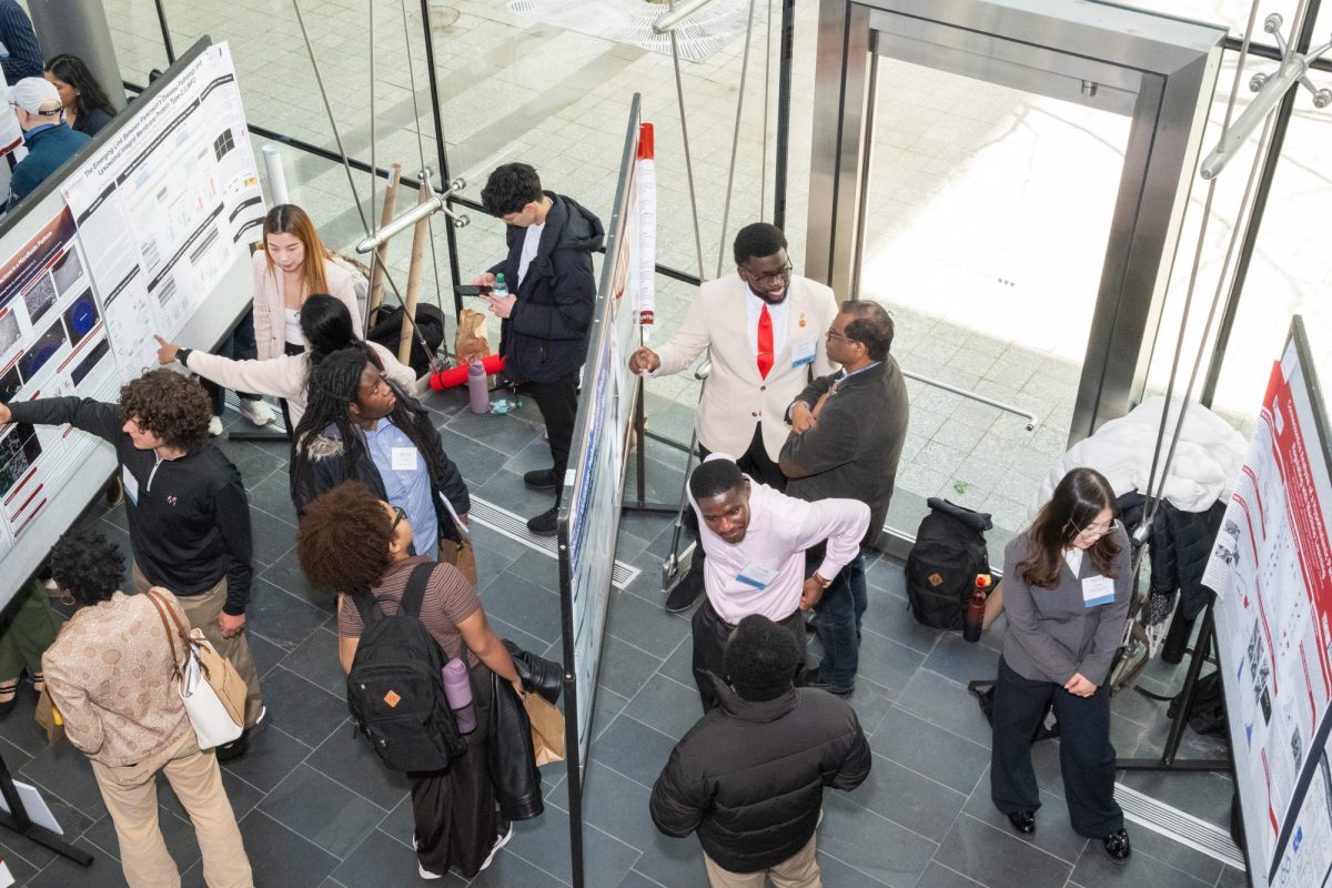
Around 50 people crowded into the Ether Dome at Massachusetts General Hospital Thursday to listen to neuroscientist Bruce Rosen and medical illustrator Danny Quirk speak about the intersectionality of human anatomy and visual art. The talk, entitled “Beautiful Brain,” was a part of HUBweek, a weeklong series of events combining art, science and technology.
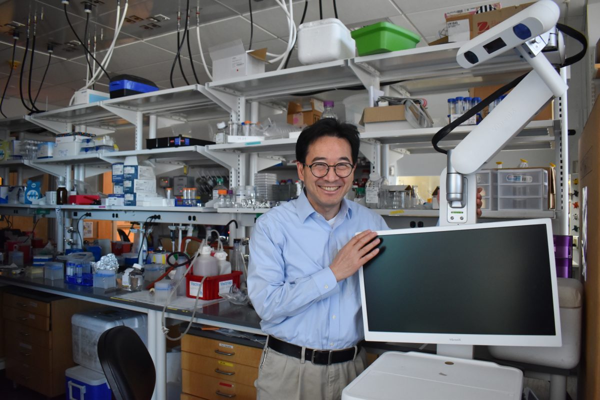
Rosen and Quirk displayed fascinating case studies and vibrant displays of the human brain to the audience. Rosen, in particular, emphasized the importance of MRI technology to his field.
“MRIs are the hub of all brain imaging,” Rosen said. “Like all good magic, it’s done with magnets.”
Rosen went on to say that since the development of the first MRI more than 30 years ago, it has been tremendously easier to identify whether a patient will go on to develop brain diseases such as Alzheimer’s later on in life. The brain imaging system now lets doctors detect small abnormalities that may have never been found before.
Rosen is currently doing extensive research to find an even more intricate method of capturing images of microscopic parts in his patients’ brains. He spoke in detail about the research that has been going on since 1995, when his colleague Van Wedeen developed a method called tractography.
“It’s a way to map connections in the human brain,” Rosen said. “Now, we can see that not only are neurons crossing, but there is branching as well.”
Rosen provided an example by describing the case of a bilingual Greek woman with ganglioglioma, a rare brain tumor. He displayed photos of the woman’s brain and pointed to parts of the brain affected by the tumor, such as those that process language comprehension.
Illuminated in green, the audience could see the areas of the woman’s brain that were responsible for her fluency in Greek. Those parts were located far from the tumor. The areas responsible for her English language skills, on the other hand, were located closer to the tumor.
“Her neurosurgeon was able to look at this image before the surgery and take extra care not to destroy the areas that processed her English language skills,” Rosen said. “There is art as well as science in these pictures. When you put it all together, you get a big bowl of spaghetti.”
Such complicated “bowls of spaghetti” are exactly what Quirk sorts through as he works. Although Quirk began as a visual artist, his early watercolors featured intricate, detailed depictions of human anatomy. Soon, he became a full-blown medical illustrator.
Quirk works closely with Rosen to develop a nuanced way to capture images of the brain. Rosen discovers the science and Quirk creates the art.
Therefore, it made sense that Quirk began his portion of the presentation by showing the audience paintings by Leonardo da Vinci and Max Brödel, the first modern medical illustrators.
“Everything Brödel did was beautifully accurate,” Quirk said.
And this is what Quirk aims for in his illustrations — the perfect balance between aesthetics and accuracy. His work has been noticed by multiple prestigious organizations, including TEDMED Live Athens and Smithsonian Magazine.
Quirk narrated his artistic journey through the world of medical illustration and the various institutions it has taken him to. He frequently visits students in universities around the country, and these institutions are so impressed by his illustrations that they incorporate body painting into the curriculum. So far, body painting has been added to the curriculum of Elon University in North Carolina and the University of Edinburgh in Scotland.
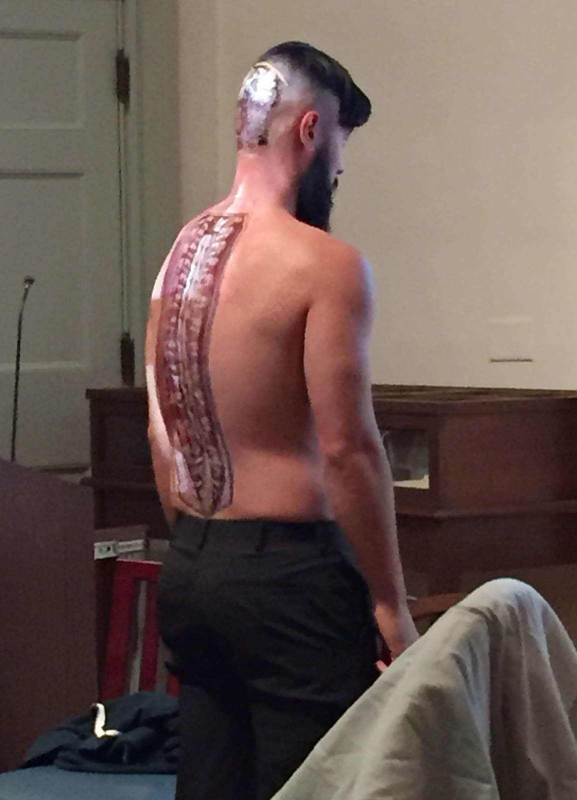
And yes, body painting is exactly what it sounds like. It involves painting cross-sections of various body parts onto the actual skin so that it gives the impression of peering straight into the flesh and marrow of a person’s body.
“It forces you to gain a deeper understanding of a specific region of the body,” Quirk said. “It turned out to be a wild success. It started off as an experiment in teaching, and then the anatomy students’ grades went up half a letter grade.”
To top off the presentation, the audience was treated with a real life depiction of a body painting. Quirk’s friend, subtly sitting in the front row of the audience, took off his sweatshirt to reveal a breathtakingly beautiful painting of the cross section of a spinal cord on his back.
The two speakers acknowledged that neuroscience research and innovative visual art techniques don’t always go hand in hand, but the unusual combination has the potential to save a patient’s life.
“We try to take these beautiful visual pictures,” Rosen said, “and use it to come up with quantitative science.”